Anatomy Rib Cage - Torso And Shoulder Anatomy Rib Cage And Abs Hd Png Download Transparent Png Image Pngitem. It is supported by the vertical sternum or breastbone (anteriorly) and the 12 thoracic vertebrae. The ribs are curved, flat bones which form the majority of the thoracic cage. Anatomy the rib cage is a bony structure found in the chest (thoracic cavity). There are twelve (12) pairs of ribs and all articulate posteriorly with the thoracic vertebrae. Each pair is numbered based on their attachment to the sternum, a bony process at the front of the rib cage which serves as an anchor point.
Related posts of muscle anatomy rib cage muscle anatomy study guide. The thoracic cage (rib cage) is the skeleton of the thoracic wall. Air reaches the lungs through the trachea, located beneath the throat. They articulate with the vertebral column posteriorly, and terminate anteriorly as cartilage (known as costal cartilage). Rib cage pain may be sharp, dull, or achy and felt at or below the chest or above the navel on either side.
Thorax Ribs Cage Anatomy Illustration 1866 High Res Vector Graphic Getty Images from media.gettyimages.com They articulate with the vertebral column posteriorly, and terminate anteriorly as cartilage (known as costal cartilage). This furrow isn't present in the 11th and 12th ribs. It is formed by the 12 thoracic vertebrae, 12 pairs of ribs and associated costal cartilages and the sternum. However, only seven have a direct articulation with the sternum. Ten of the twelve ribs connect to strips of hyaline cartilage on the anterior side of the body. The thoracic cage (rib cage) forms the thorax (chest) portion of the body. It is supported by the vertical sternum or breastbone (anteriorly) and the 12 thoracic vertebrae. Vintage anatomy print features the human rib cage and shoulders.
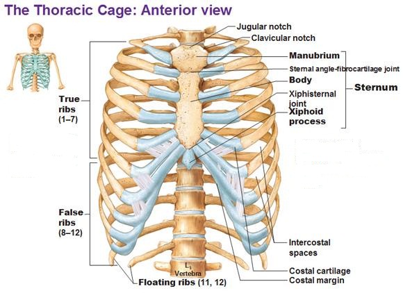
Anatomy of the rib cage diagram in this image, you will find thoracic vertebrum, costochondral joint, costal cartilage, costal margin, costal arch, thoracic vertebrum, xiphoid process, xiphisternal joint, body, manubrial sternal joint, manubrium, the sternal notch in it.
Anatomy of human stomach 10 photos of the anatomy of human stomach anatomy human colon, anatomy human digestive system, anatomy human heart, anatomy human kidney, anatomy human liver, anatomy human pancreas, anatomy human spleen, human body stomach, stomach, anatomy human colon, anatomy human digestive system, anatomy. There are 11 pairs of external intercostal muscles. The cartilage strips are called costal cartilage (costal is the anatomical adjective that refers to the rib) and connect on their other end to the sternum. Vintage anatomy print features the human rib cage and shoulders. On the interior wall of the rib body is a channel, sulcus costae, with blood vessels and nerves. Rib cage pain may be sharp, dull, or achy and felt at or below the chest or above the navel on either side. Anatomy of the rib cage diagram in this image, you will find thoracic vertebrum, costochondral joint, costal cartilage, costal margin, costal arch, thoracic vertebrum, xiphoid process, xiphisternal joint, body, manubrial sternal joint, manubrium, the sternal notch in it. The lungs are two separate but connected organs located in the upper chest, covered by the rib cage. The upper edge is round and the lower sharp. Lateral view of a pair of ribs articulating with the thoracic vertebrae. There are twelve pairs of ribs, all of which articulate with the vertebral column. It is formed by the 12 thoracic vertebrae, 12 pairs of ribs and associated costal cartilages and the sternum. Introduction to the structure of the ribcage and ribs:
In this episode we'll learn about the simple structure of the rib cage and have a look at the detailed anatomical parts of the ribs. The cartilage strips are called costal cartilage (costal is the anatomical adjective that refers to the rib) and connect on their other end to the sternum. However, only seven have a direct articulation with the sternum. The rib below that is rib 2, and it connects to the t2 thoracic vertebra, and so on. The ribs are a veritable collection of bone, muscle, and organs, most of which are fairly important for living and other useful functions.
How Many Ribs Does The Human Body Have What Are False Ribs Socratic from useruploads.socratic.org It is formed by the 12 thoracic vertebrae, 12 pairs of ribs and associated costal cartilages and the sternum. Vintage anatomy print features the human rib cage and shoulders. Air reaches the lungs through the trachea, located beneath the throat. Each pair is numbered based on their attachment to the sternum, a bony process at the front of the rib cage which serves as an anchor point. The ribs are a set of twelve paired bones which form the protective 'cage' of the thorax. 5 out of 5 stars (81) $ 16.71. The thoracic cage takes the form of a domed bird cage with the horizontal bars formed by ribs and costal cartilages. Rib cage anatomy the rib cage, shaped in a mild cone shape and more flexible than most bone sets, is made up of varying elements such as the thoracic vertebra, 12 equally paired ribs, costal cartilage, and held together anteriorly by the sternum.
The thoracic cage (rib cage) forms the thorax (chest) portion of the body.
It is formed by the 12 thoracic vertebrae, 12 pairs of ribs and associated costal cartilages and the sternum. The lungs are two separate but connected organs located in the upper chest, covered by the rib cage. It consists of the 12 pairs of ribs with their costal cartilages and the sternum (figure 6.38). It may occur after an obvious injury or without explanation. The top edge of the manubrium has a depression called the suprasternal or jugular notch. The thoracic cage consists of the 12 thoracic vertebrae, the associated intervertebral discs, 12 pairs of ribs with their costal cartilages, and the sternum. As part of the bony thorax, the ribs protect the internal thoracic organs. On the interior wall of the rib body is a channel, sulcus costae, with blood vessels and nerves. Rib cage pain may be sharp, dull, or achy and felt at or below the chest or above the navel on either side. This furrow isn't present in the 11th and 12th ribs. The cartilage strips are called costal cartilage (costal is the anatomical adjective that refers to the rib) and connect on their other end to the sternum. The thoracic cage protects the heart and lungs. In this video, we explore:1) the anatomy of the sternum2) the anatomy and differences between the three classes of ribs3) the anatomy and differences between.
Ten of the twelve ribs connect to strips of hyaline cartilage on the anterior side of the body. This furrow isn't present in the 11th and 12th ribs. Pain under the left rib cage can mean anything from a ruptured spleen, to heart trouble, to just needing to have a good fart. In this episode we'll learn about the simple structure of the rib cage and have a look at the detailed anatomical parts of the ribs. The sternum is a flat bone that is made up of three parts, the (1) manubrium, (2) body, and the (3) xiphoid process.
Anatomy Of The Rib Cage Diagram from www.anatomynote.com The rib below that is rib 2, and it connects to the t2 thoracic vertebra, and so on. In this video, we explore:1) the anatomy of the sternum2) the anatomy and differences between the three classes of ribs3) the anatomy and differences between. The thoracic cage takes the form of a domed bird cage with the horizontal bars formed by ribs and costal cartilages. It is made up of 12 pairs of ribs. As part of the bony thorax, the ribs protect the internal thoracic organs. Anatomy the rib cage is a bony structure found in the chest (thoracic cavity). Each pair is numbered based on their attachment to the sternum, a bony process at the front of the rib cage which serves as an anchor point. Pain under the left rib cage can mean anything from a ruptured spleen, to heart trouble, to just needing to have a good fart.
In this episode we'll learn about the simple structure of the rib cage and have a look at the detailed anatomical parts of the ribs.
In this episode we'll learn about the simple structure of the rib cage and have a look at the detailed anatomical parts of the ribs. It may occur after an obvious injury or without explanation. The lungs are two separate but connected organs located in the upper chest, covered by the rib cage. However, only seven have a direct articulation with the sternum. Contributing to their role in protecting the internal thoracic organs. There are twelve (12) pairs of ribs and all articulate posteriorly with the thoracic vertebrae. On the interior wall of the rib body is a channel, sulcus costae, with blood vessels and nerves. Pain under the left rib cage can mean anything from a ruptured spleen, to heart trouble, to just needing to have a good fart. The thoracic cage (rib cage) is the skeleton of the thoracic wall. At the chest, many rib bones connect to the sternum via costal cartilage,. There are 11 pairs of external intercostal muscles. It is supported by the vertical sternum or breastbone (anteriorly) and the 12 thoracic vertebrae. The thoracic cage (rib cage) forms the thorax (chest) portion of the body.
Share :
Post a Comment
for "Anatomy Rib Cage - Torso And Shoulder Anatomy Rib Cage And Abs Hd Png Download Transparent Png Image Pngitem"


Post a Comment for "Anatomy Rib Cage - Torso And Shoulder Anatomy Rib Cage And Abs Hd Png Download Transparent Png Image Pngitem"